Home
/ Back Of Skull Anatomy - Ronten Human Skull Model Life Size Replica Medical Anatomy Anatomical Adult Model With Removable Skull Cap And Articulated Mandible Full Set Of Teeth 7 2x4 2x4 95in Amazon Com Industrial Scientific
Back Of Skull Anatomy - Ronten Human Skull Model Life Size Replica Medical Anatomy Anatomical Adult Model With Removable Skull Cap And Articulated Mandible Full Set Of Teeth 7 2x4 2x4 95in Amazon Com Industrial Scientific
Back Of Skull Anatomy - Ronten Human Skull Model Life Size Replica Medical Anatomy Anatomical Adult Model With Removable Skull Cap And Articulated Mandible Full Set Of Teeth 7 2x4 2x4 95in Amazon Com Industrial Scientific. It is comprised of many bones, formed by intramembranous ossification, which are joined together by sutures (fibrous joints). Learn about the anatomy of the skull bones and sutures as seen on ct images of the brain. First, the lambdoid suture connects the occipital bone to both parietal bones. Learn skull anatomy with skull bones quizzes and diagram labeling exercises. Hank grebe / getty images
It can be easily felt through the. In the adult, the skull consists of 22 individual bones, 21 of which are immobile and united into a single unit. A bump on the back of the head has many possible causes, including injuries, cysts, fatty growths, inflamed hair follicles, and bone spurs. See human skull anatomy stock video clips. It is formed by a chain of 33 interconnected vertebrae and their intervening joints.
Understanding Skull Base Tumors University Hospitals from api.kramesstaywell.com The bone that rests on top of your spine the occipital bone is a bone that covers the back of your head; It is also known as the calvarium. A bump on the back of the head has many possible causes, including injuries, cysts, fatty growths, inflamed hair follicles, and bone spurs. The erector spinae are a group of many muscles that attach along the back of the spine. The foramen magnum, housing the brainstem, is also a part of the occipital bone. This usually stems from tension in the muscles in the neck. In order to be light, the skull is made up by flat and irregular bones, and has hollow spaces called the sinuses. It offers protection to the brain, eye balls, inner ears, and nasal passages.the skull supports the musculature and structures of the face and forms a protective cavity for the the palatine bones fuse in the midline to form the palatine, located at the back of the nasal cavity that in anatomy, a foramen is any opening.
There are several important features to know about the occipital bone.
In order to be light, the skull is made up by flat and irregular bones, and has hollow spaces called the sinuses. The occipital bone overlies the occipital lobes of the cerebrum. This portion of the skull base consists of the orbital portion of the frontal bone. We use cookies to ensure that we give you the best experience on our website. Related posts of bone of back of skull bones and muscles labeled. The occipital bone surrounds a large opening known as the foramen magnum. The skull is a strong, bony capsule that rests on the neck and encloses the brain. The foramen magnum, housing the brainstem, is also a part of the occipital bone. The occipital bone is the only bone in your head that connects with your cervical spine (neck). As a review activity, label figures 131, 132, 13 3, 134, and 135 back of skull anatomy. The occipital bone houses the back part of the brain and is one of seven bones that come together to form the skull. They are attached at the back of the head, and usually give rise for a headache. See anatomy of the head and neck stock video clips.
The occipital bone is located at the back of the skull and protects the underlying cerebellum, brainstem, and occipital lobe of the cerebrum. It is located next to five of the cranium bones. The bone that rests on top of your spine the occipital bone is a bone that covers the back of your head; Bumps on this part of the body can be hard or soft, and. 24 years experience orthopedic spine surgery.
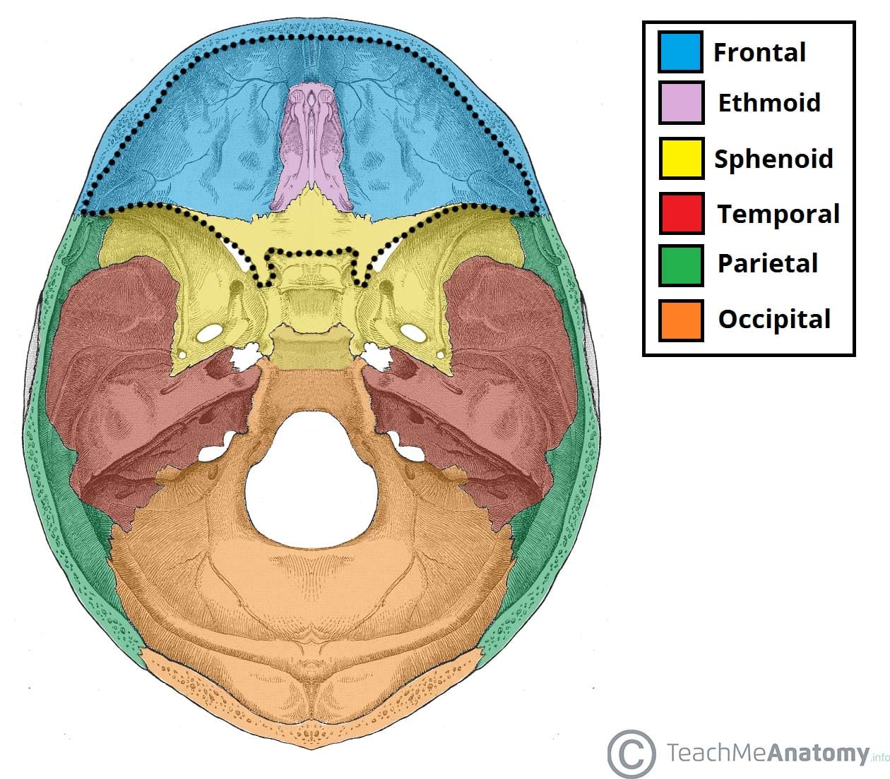
Anterior Cranial Fossa Boundaries Contents Teachmeanatomy from teachmeanatomy.info Frontal, sphenoid, ethmoid, occipital, parietal and temporal. We use cookies to ensure that we give you the best experience on our website. Learn about the anatomy of the skull bones and sutures as seen on ct images of the brain. The skull has a single occipital condyle.7 the skull consists of five major bones: The occipital bone is the only bone in your head that connects with your cervical spine (neck). The occipital bone overlies the occipital lobes of the cerebrum. The occipital bone houses the back part of the brain and is one of seven bones that come together to form the skull. The ethmoid bone forms the central part of the floor, which is the deepest area of the anterior cranial fossa.
The skull is a strong, bony capsule that rests on the neck and encloses the brain.
Learn about the anatomy of the skull bones and sutures as seen on ct images of the brain. Hank grebe / getty images The occipital bone (/ ˌɒkˈsɪpɪtəl /) is a cranial dermal bone and the main bone of the occiput (back and lower part of the skull). The mayo clinic says that these factors can be chemical activity in your brain, the nerves of blood vessels surrounding your skull, or the muscles at the back of the head and neck. The pain in the back of your head occurs when one or a combination of factors affect your brain. It is trapezoidal in shape and curved on itself like a shallow dish. The ethmoid bone forms the central part of the floor, which is the deepest area of the anterior cranial fossa. Bones and muscles labeled 12 photos of the bones and muscles labeled bone and muscle labeling quiz, bones and muscles labeled, bone, bone and muscle labeling quiz, bones and muscles labeled They are attached at the back of the head, and usually give rise for a headache. It forms the axial skeleton together with the skull and rib cage. The vertebral column (spine) is the bony core of the back. The skull is the bony skeleton of the head. 1 also, some people are more prone to headaches than others.
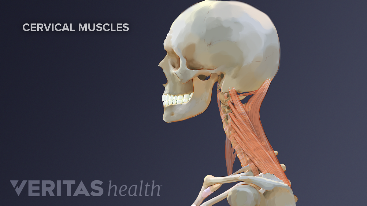
Axial muscles of the head, neck, and back. The pain in the back of your head occurs when one or a combination of factors affect your brain. Bones and muscles labeled 12 photos of the bones and muscles labeled bone and muscle labeling quiz, bones and muscles labeled, bone, bone and muscle labeling quiz, bones and muscles labeled It can be easily felt through the. The occipital bone is located at the back of the skull and protects the underlying cerebellum, brainstem, and occipital lobe of the cerebrum.
Neck Muscles And Other Soft Tissues from embed.widencdn.net Massaging the sinus pressure points at the back of the head. An area called the occiput. Hank grebe / getty images This portion of the skull base consists of the orbital portion of the frontal bone. The occipital bone is the small bony protrusion at the base of the skull where the skull joins to the neck. It forms the axial skeleton together with the skull and rib cage. It is trapezoidal in shape and curved on itself like a shallow dish. It consists of two major parts:
In order to be light, the skull is made up by flat and irregular bones, and has hollow spaces called the sinuses.
It is also known as the calvarium. We use cookies to ensure that we give you the best experience on our website. Massaging the sinus pressure points at the back of the head. Muscle head anatomy vocal organ diagram female neck anatomy neck wireframe head neck human anatomy head artery anatomy face pharynx vector neck degree head anatomy 3d. Learn about the anatomy of the skull bones and sutures as seen on ct images of the brain. The greater portion of the anterior floor is convex and grooved by the frontal lobe gyri. Bones of cranium there are eight major bones and eight auxiliary bones of the cranium. It is comprised of many bones, formed by intramembranous ossification, which are joined together by sutures (fibrous joints). The occipital bone is the only bone in your head that connects with your cervical spine (neck). The erector spinae are a group of many muscles that attach along the back of the spine. It is made up of more than 100 billion nerves that communicate in trillions of connections called synapses. The occipital bone surrounds a large opening known as the foramen magnum. A thorough description is beyond the.
Share :
Post a Comment
for "Back Of Skull Anatomy - Ronten Human Skull Model Life Size Replica Medical Anatomy Anatomical Adult Model With Removable Skull Cap And Articulated Mandible Full Set Of Teeth 7 2x4 2x4 95in Amazon Com Industrial Scientific"

Post a Comment for "Back Of Skull Anatomy - Ronten Human Skull Model Life Size Replica Medical Anatomy Anatomical Adult Model With Removable Skull Cap And Articulated Mandible Full Set Of Teeth 7 2x4 2x4 95in Amazon Com Industrial Scientific"